


Monitoring brain tsunamis with graphene transistors
Graphene devices can transform neurological care
Just as tsunamis are massive waves that surge across the ocean, waves of electrical disturbances can sweep through the brain and disrupt its normal functioning. In technical terms these abnormal brain activities are known as spreading depolarizations and are triggered in traumatic brain injuries, strokes and other neurological diseases.
What happens during a brain tsunamis?
The brain looks like a complex tapestry of neurons that communicate with each other using both a chemical and an electrical language. Each neuron responds to a balanced cocktail of chemical and electrical signals coming from neighbouring neurons. During spreading depolarization, this “cocktail” is disrupted, and the neurons become less able to function properly. It's comparable to how a tsunami can disrupt the normal functioning of the regions it hits.
Spreading depolarizations have a very low frequency below 0.1 Hz, meaning that they spread slowly. The frequency is significantly lower compared to gamma waves (typical of information processing, 30 Hz), beta waves (problem solving, 14-30 Hz), alpha waves (wakeful relaxation, 8-13 Hz), theta waves (light sleep, 4-8 Hz) and delta waves (deep sleep, 0.5-4 Hz). Research on spreading depolarizations is still ongoing, and its precise functions and implications are not yet fully understood.
How can brain tsunamis be recorded?
Glass microelectrodes, also called pipettes, have long been a fundamental tool in the field of electrophysiology. However, they are limited to measure brain electrical signals in one or a few points. To cover a larger area of the brain, Graphene Flagship researchers have developed flexible graphene field-effect transistors (FETs) and depth neural probes. They have used graphene depth neural probes to study the brain activity of awake rodents suffering from epilepsy. They tested the long-term functionality of these devices by implanting them in rats for 10 weeks. During this time, they were able to record patterns of brain activity seen in certain types of epilepsy and the associated slow brain waves (infraslow oscillations).
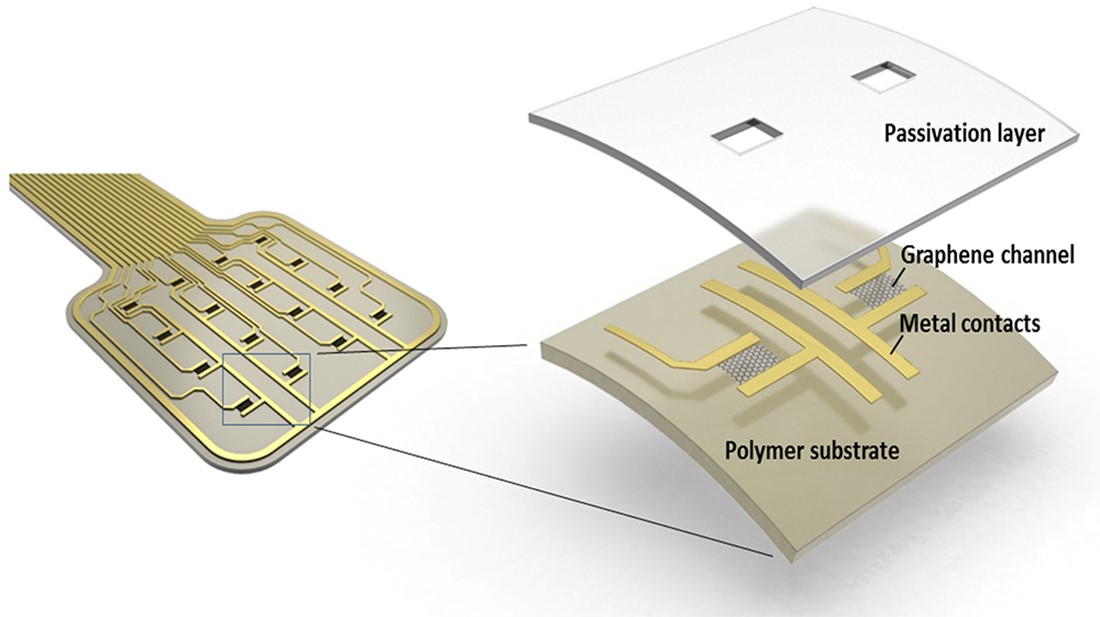
Probe layout (Credit: Eduard Masvidal-Codina)
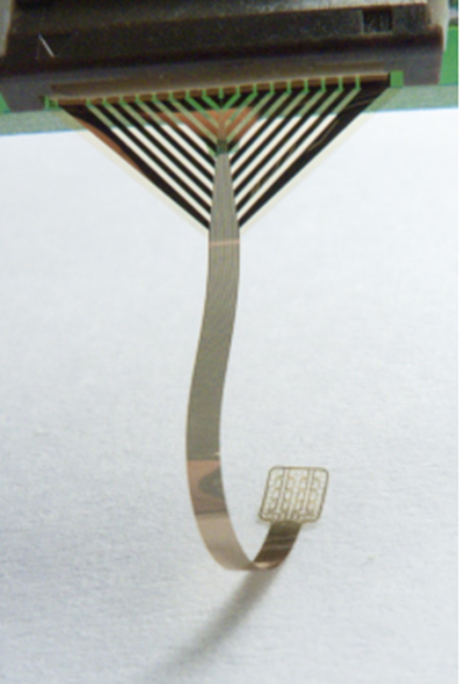
Flexible neural probe to record electrical signals from the brain. (Credit: Nature Materials)
In the video you can watch Eduard Masvidal, researcher at ICN2 (Spain), who has been working on graphene-FET devices for neurophysiology and the monitoring of brain tsunamis as a member of the Graphene Flagship Work Package on Biomedical Technologies.
References
Masvidal-Codina, Eduard, et al. "Characterization of optogenetically-induced cortical spreading depression in awake mice using graphene micro-transistor arrays." Journal of Neural Engineering 18.5 (2021): 055002. https://iopscience.iop.org/article/10.1088/1741-2552/abecf3/meta
Bonaccini Calia, Andrea, et al. "Full-bandwidth electrophysiology of seizures and epileptiform activity enabled by flexible graphene microtransistor depth neural probes." Nature Nanotechnology 17.3 (2022): 301-309. https://www.nature.com/articles/s41565-021-01041-9
Masvidal-Codina, Eduard, et al. "High-resolution mapping of infraslow cortical brain activity enabled by graphene microtransistors." Nature materials 18.3 (2019): 280-288. https://www.nature.com/articles/s41563-018-0249-4
Author bio

Letizia Diamante
Science Writer and Coordinator of the 'Diversity in Graphene' initiative.