Transforming cancer diagnosis
A turnkey instrument to support clinicians and histopathologists
Graphene Flagship Partner Cambridge Raman Imaging (CRI) is developing a technology to diagnose cancer quickly and at affordable costs. The instrument measures the molecular composition of tissue biopsies using coherent Raman – a microscopy technique that differentiates molecules by the way they vibrate, and is three - five orders of magnitude quicker than spontaneous Raman.
CRI was founded in 2018 in Cambridge (UK), as a spin-out of the University of Cambridge and Politecnico di Milano (Italy). This year CRI was selected to coordinate CHARM, a €3.3 million grant of the European Innovation Council’s Transition call, and raised £1.1 million through equity funding. Our discussion with Matteo Negro, CRI’s CEO and CTO follows.
Which problem in cancer diagnosis are you trying to solve?
Cancer is one of the biggest burdens in our society, but hospitals are currently under increasing pressure due to a large number of cancer cases and the reduction of medical personnel. The current routine for tumour diagnosis is subjective and time consuming, because it is based on an old staining process of preparing biopsy samples. Cambridge Raman Imaging is developing an innovative laser source based on graphene related materials that provides an objective signature of the presence or the absence of tumours directly from the unstained tissue samples.
Can you explain what you mean by objective signature?
Our Coherent Raman Imaging technology allows us to extract Raman spectra in every single point of the sample in a few seconds or minutes. It is the first application of fast Raman imaging to the clinical field. The Raman measurements are then directly fed into an Artificial Intelligence (AI) programme that differentiates healthy and tumour tissues, and provides tumour subtype classification. The pathologists will not need to produce a negative control for every patient, and can spend more time analysing the most critical cases.
What’s your final goal?
We aim to develop a medical device that is capable of automatically analysing unstained tissues, differentiating normal versus neoplastic tissues with accuracy higher than 98%, and differentiating and grading histologic subtypes with accuracy higher than 90%. Our instrument will offer an accuracy comparable to the existing clinical protocols, but it will be much faster and more objective. Our diagnostic medical device will support clinicians and histopathologists, and we are already collaborating with them.
Can this technology find other applications beyond cancer?
Yes, the extreme chemical sensitivity and fast imaging capabilities of this technology can be exploited for a really large number of different applications. Beyond the biomedical and clinical applications, our technology can be successfully applied to the study of food contamination and environmental pollutants, like microplastics. This technology can also be used to monitor the degradation of batteries.

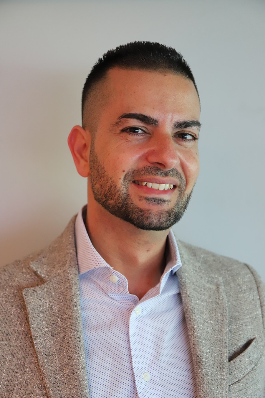
Matteo Negro, CRI’s CEO and CTO


